

Why a Plucked Hair Follicle May Matter to the Future of Regenerative Medicine
In this article, I return to a study that I discussed in a July Journal Club post. The study, published in Cells in 2024, investigated whether cells obtained from plucked, cryopreserved human hair follicles could be reprogrammed into induced pluripotent stem cells and then directed toward a pancreatic lineage. The practical goal was to develop a non-invasive, patient-specific source of cells that might eventually support disease modeling or autologous cell-replacement strategies for type 1 diabetes.
As you know, when I review a paper for Journal Club, I always want to begin by placing it at the correct level of evidence. That is especially important in regenerative medicine, where exciting laboratory findings can quickly be described as though they are already treatments.
This study is not a cure for type 1 diabetes. It is not a human treatment trial, and it does not report changes in A1C, insulin requirements, hypoglycemia, or any other clinical outcome. It is an early-stage, preclinical study showing that cells recovered from cryopreserved, plucked human hair follicles can be reprogrammed into induced pluripotent stem cells, or iPSCs, and then directed toward a pancreatic progenitor lineage.
That may sound like a narrow laboratory achievement, but through the lens of Cellular Medicine, it represents an important advance. The significance is not simply that the researchers produced cells carrying pancreatic markers. The larger advance is that they created a practical workflow beginning with an easily accessible, non-invasive source of a patient’s own cells.
Beginning With the Right Cell
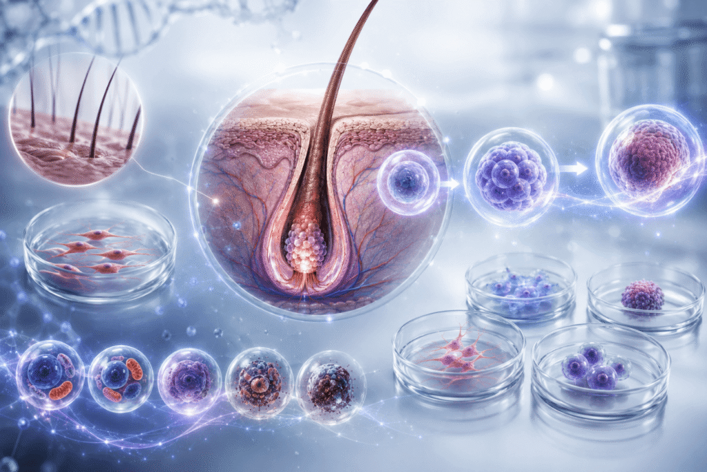
For years, researchers have been able to take a mature adult cell, which is often a fibroblast obtained through a skin biopsy, and reprogram it into an induced pluripotent stem cell. This process uses a group of reprogramming factors associated with the pioneering work of Shinya Yamanaka. These factors effectively turn back portions of the cell’s developmental program, returning a specialized adult cell to a more primitive state from which it can potentially be directed into another lineage.
In this paper, the investigators asked whether we might begin somewhere more accessible than a skin biopsy: the hair follicle.
A hair follicle is not simply a strand of hair. It is more like a miniature regenerative ecosystem. It contains keratinocytes, fibroblasts, dermal papilla cells, stromal cells, and specialized stem-cell niches within regions such as the follicular bulge.
That diversity matters. Some cells within the follicle may be more developmentally flexible or more readily reprogrammable than a conventional adult fibroblast. They may require less biological work to return to a pluripotent state. This particular study does not prove that mechanism, but it raises an important possibility: the tissue source we select may have a major effect on the efficiency and quality of the regenerative process that follows.
The starting cell is not a neutral ingredient. Its age, metabolic health, mitochondrial integrity, accumulated DNA damage, epigenetic state, degree of senescence, and ability to respond to growth signals can all influence what happens next. In Cellular Medicine, we are always interested in that starting condition because the future behavior of a cell is shaped by the biological history it carries with it.
What the Researchers Actually Did
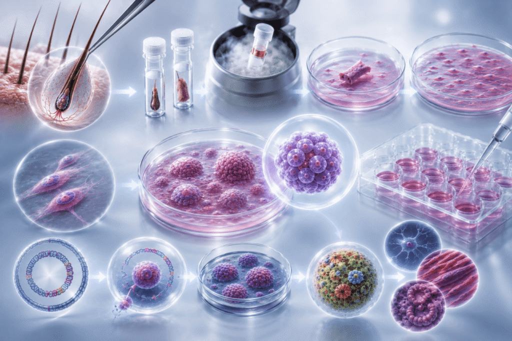
The investigators collected hair follicles from the occipital region of the scalp in 28 adult participants of different ages and both sexes. The follicles were transported, cryopreserved, thawed, and placed into an explant culture system. The researchers selected follicles with an intact outer root sheath and allowed cells to grow outward from the follicular tissue.
They were then able to isolate and expand keratinocytes, identified through markers such as K14 and K5. In some cases, a single successful follicular outgrowth produced approximately 1.2 million cells by the second passage—enough material to proceed with cellular reprogramming.
The next step was to take keratinocytes from 11 of the participants and expose them to a non-viral, integration-free episomal reprogramming system. The vectors carried several familiar reprogramming factors, including OCT4, SOX2, KLF4, L-MYC, and LIN28. The use of an integration-free system is important because it reduces the concern that the reprogramming material will permanently insert itself into the cellular genome.
By day 21, every one of the 11 participant samples had produced at least one iPSC colony. Next, four of those cell lines were examined more extensively. The researchers selected two lines from individuals younger than 40 and two from individuals older than 60. These cells expressed established pluripotency markers, including OCT4, NANOG, and SOX2. Over serial passages, the episomal vectors were cleared, and conventional karyotyping did not identify major chromosomal abnormalities in the four lines tested.
The researchers also tested whether the iPSCs could differentiate into the three major embryonic germ layers: ectoderm, mesoderm, and endoderm. They demonstrated this both in culture and through a teratoma assay in immunodeficient mice. The mouse assay was not a treatment experiment. It was used to establish that the cells retained pluripotency—the ability to produce tissues representing all three germ layers.
Directing the Cells Toward the Pancreas
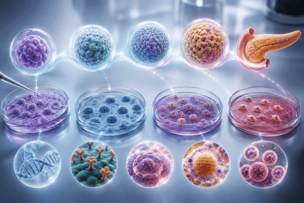
Once the investigators had validated the iPSC lines, they directed them toward definitive endoderm. This is an important developmental stage because the pancreas arises from the endodermal germ layer.
The differentiated cells expressed SOX17 and FOXA2, both associated with definitive endoderm. Between approximately 95.6 and 97.9 percent of the cells also co-expressed the surface markers cKIT and CXCR4.
The researchers then carried one cell line further through a modified differentiation protocol. At the end of that process, the cells expressed PDX1 and NKX6.1. These are significant markers because their co-expression is associated with pancreatic progenitor identity.
And this is where I think we need to be very precise. These were not mature pancreatic beta cells. They did not demonstrate glucose-stimulated insulin secretion. They did not show C-peptide production, restoration of normal glucose levels, or insulin independence. The investigators showed that the cells had reached a pancreatic progenitor-like stage from which further beta-cell development may be possible.
That distinction is not a criticism of the paper. It is simply an accurate description of what was accomplished.
A progenitor cell is a cell moving in the correct developmental direction. It has acquired part of the identity needed to become a more specialized cell, but it has not necessarily reached functional maturity. Marker expression tells us that the developmental program is moving toward the pancreas. It does not yet tell us that the cell can perform the integrated work of a beta cell.
Identity Is Not the Same as Function
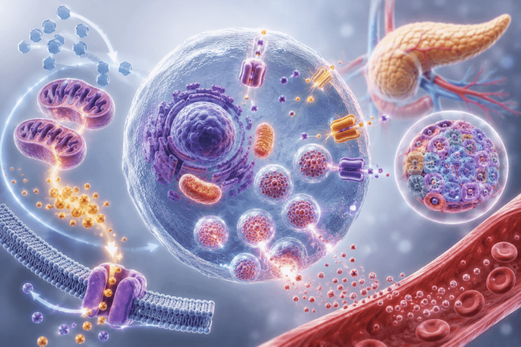
A mature beta cell must do far more than express pancreatic markers. It must sense changes in circulating glucose, metabolize that glucose, couple changes in ATP production to membrane ion channels, generate calcium signaling, and release insulin in a controlled and physiologically appropriate manner.
That entire sequence depends on cellular energetics. Glucose must be processed through glycolysis and mitochondrial oxidative metabolism. The rise in the ATP-to-ADP ratio must influence ATP-sensitive potassium channels. Membrane depolarization must open calcium channels, and calcium entry must trigger insulin granule exocytosis. The cell must produce, package, store, and release insulin at the right time and in the right amount.
Significantly, none of those functional steps was tested in this study.
The next stage of research must determine whether these pancreatic progenitor cells can mature into cells that produce insulin and C-peptide, respond dynamically to glucose, remain stable over time, survive transplantation, establish adequate vascular support, and improve glucose control in an appropriate diabetic animal model.
In my Journal Club discussion, I highlighted immune activation, graft survival, C-peptide production, and glucose-stimulated insulin secretion as essential future endpoints. Those functional tests are where we move from saying, “This cell has the right developmental markers,” to saying, “This cell can actually perform the job we need it to do.”
Why the Cell Source Is So Important
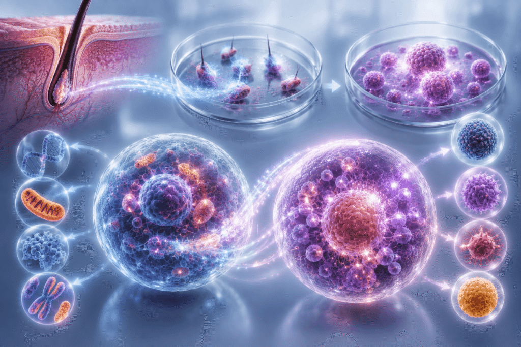
One of the most interesting findings in the paper was the relationship between follicular outgrowth and reprogramming efficiency. Samples that produced stronger cellular outgrowth also tended to generate more iPSC colonies.
That tells us something very important: the quality of the starting material matters. Even when researchers begin with the same number of cells and use the same reprogramming factors, the cells do not all respond equally. Some may be biologically primed to proliferate and reprogram more efficiently. The authors discuss the possibility that certain subpopulations possess what has been called reprogramming “eliteness”—a selective biological advantage that allows them to transition into the iPSC state more readily.
From an SSRP perspective, this opens a much broader set of questions. What makes one person’s cells more viable or reprogrammable than another’s? Is the difference related to mitochondrial function, oxidative stress, DNA repair, inflammatory signaling, nutrient sensing, senescence, or epigenetic age? Can the biological quality of the starting cell be improved before reprogramming? Can we identify the most useful follicular subpopulation before beginning the process?
The paper does not answer those questions, but it gives us a practical model through which they can now be studied.
The Possibility of Autologous Cell Banking
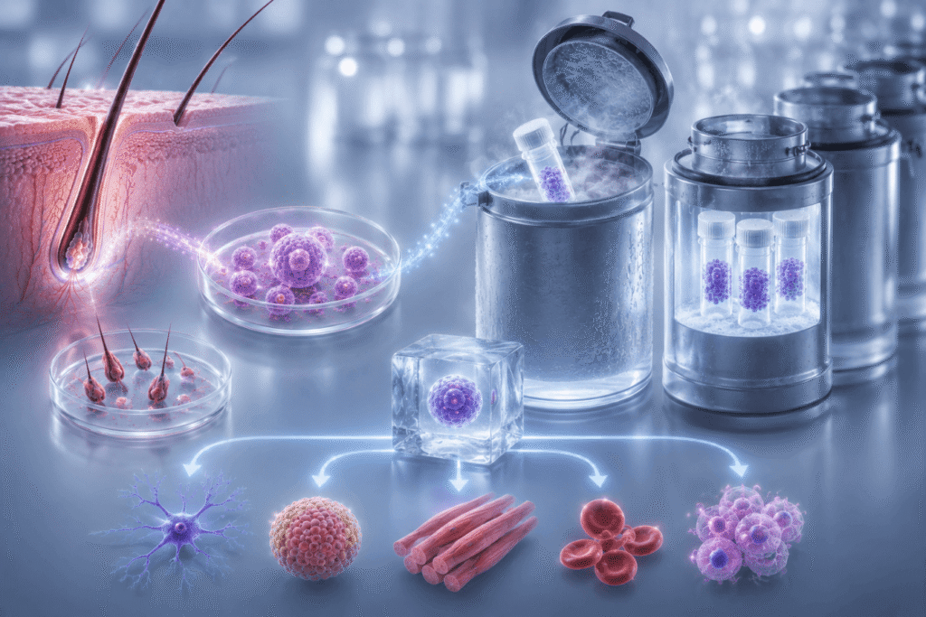
The practical nature of hair follicle collection may be the most consequential aspect of this research. A skin biopsy requires a medical procedure. Bone marrow collection is considerably more invasive. Umbilical cord tissue is available only at birth. Hair follicles, by contrast, can be collected without surgery, transported, cryopreserved, and potentially banked for later use.
That creates the possibility of preserving a person’s cells earlier in life, before they accumulate additional age-related damage. I am not suggesting that this is already a proven clinical strategy. We do not yet know whether banking hair follicles will reliably produce future therapies, and many technical, economic, regulatory, and safety questions remain.
But the logic deserves serious consideration.
As cells age, they may accumulate nuclear and mitochondrial DNA mutations, epigenetic drift, oxidative injury, protein damage, and diminished repair capacity. Reprogramming can reset some features associated with cellular age, but it cannot be assumed to erase every mutation or defect present in the original cell.
The paper itself notes that donor-specific mutations can persist through iPSC generation and that the evidence examining the effects of donor age remains limited by small numbers of studied cell lines.
A younger stored cell may therefore provide a more favorable starting point than a cell collected decades later. That remains a hypothesis requiring long-term validation, but it illustrates why accessible autologous cell banking could become an important part of regenerative medicine.
Autologous Does Not Mean Immune-Proof
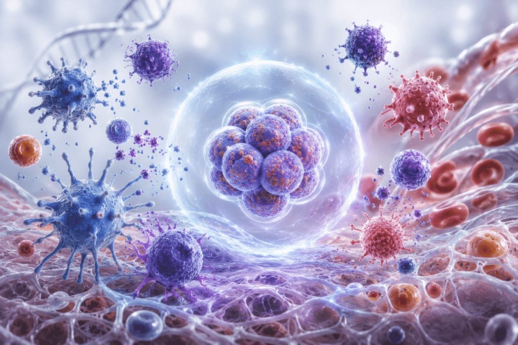
The fact that these cells originate from the patient is another major advantage. Autologous cells are genetically matched to the recipient and may reduce the risk of rejection caused by donor-recipient mismatch.
However, type 1 diabetes presents an additional problem. It is an autoimmune disease. The immune system has already demonstrated the ability to recognize and destroy insulin-producing beta cells.
Therefore, even if we generate genetically matched beta cells from a patient’s own hair follicles, we cannot assume that the autoimmune process will ignore them. Autologous sourcing may reduce alloimmune rejection, but it does not necessarily eliminate recurrent autoimmunity.
A durable treatment may ultimately require several coordinated components: beta-cell replacement, immune modulation, restoration of immune tolerance, graft protection, vascular integration, and support of the extracellular and metabolic environment surrounding the transplanted cells.
This is why I continually emphasize that a cell cannot be understood in isolation. Its survival and behavior depend on the signals it receives from the immune system, the circulation, the extracellular matrix, neighboring cells, and the metabolic environment.
Safety Must Remain Central
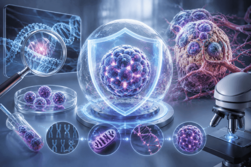
The ability to create pluripotent cells comes with real safety concerns. A pluripotent cell can generate many tissue types. That is its therapeutic promise, but it is also why uncontrolled or residual pluripotent cells can form teratomas.
The researchers intentionally used a teratoma assay in mice to prove that the iPSC lines could generate tissues from all three germ layers. Before any clinical application, however, residual pluripotent cells would have to be removed with a very high degree of confidence.
Genomic stability must also be evaluated more deeply. The normal karyotypes reported in four lines are encouraging, but standard karyotyping detects only relatively large chromosomal abnormalities. It does not rule out smaller mutations, mitochondrial DNA changes, epigenetic abnormalities, or rare subclones within a cell population.
I noted in the Journal Club discussion that genomic instability, mitochondrial mutations, residual pluripotent cells, and teratoma formation all require further validation.
These concerns do not diminish the significance of the study. They define the safety work that must accompany its continued development.
Why I Believe This Paper Matters
The most useful way to understand this research is as a demonstrated sequence:
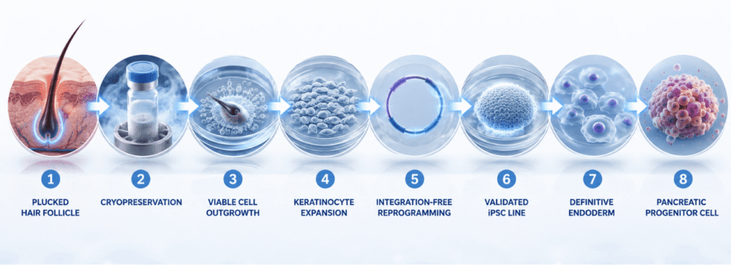
That is a meaningful achievement.
The researchers did not show that these cells cure diabetes. They showed that a tissue we can obtain easily and non-invasively may serve as a reliable, patient-specific starting point for building pancreatic cell models and, perhaps one day, replacement cells.
The same platform may also be useful well beyond the pancreas. Once a stable patient-specific iPSC line has been generated, it can potentially be directed toward neural, cardiac, hepatic, musculoskeletal, or other cellular lineages. It may be used to study disease mechanisms, screen drugs, or test how an individual’s cells respond to particular biological stresses.
This is why I view the paper as opening a door. It does not take us through every step required for a clinical therapy, but it makes the first steps easier, more accessible, and potentially more personalized.
The authors themselves acknowledge important limitations. Only four iPSC lines underwent extensive characterization, and pancreatic progenitor differentiation was performed in a limited experiment. There were no functional insulin studies and no demonstration of therapeutic efficacy. As I said in the Journal Club discussion, it is a major clinical leap to describe these as functional beta cells. They are pancreatic progenitor cells carrying markers that give us reason to believe they may be moved toward that next stage.
For me, the larger Cellular Medicine lesson is this: the body contains accessible reservoirs of biological potential. Our task is to learn how to preserve that potential, evaluate its quality, reprogram it safely, and direct it with enough precision to restore function.
The hair follicle may prove to be an important tool in that process. Not because a hair follicle is itself a pancreatic cell, but because it contains living, patient-specific cells that can be redirected toward an entirely different biological purpose.
That is not yet a treatment for type 1 diabetes. But it is exactly the kind of foundational work from which future treatments are built.
Reference:
Fatehi, A., Sadat, M., Fayyad, M., Tang, J., Han, D., Rogers, I. M., & Taylor, D. (2024). Efficient Generation of Pancreatic Progenitor Cells from Induced Pluripotent Stem Cells Derived from a Non-Invasive and Accessible Tissue Source-The Plucked Hair Follicle. Cells, 13(12), 1010. https://doi.org/10.3390/cells13121010