

Mitochondrial dysfunction is one of the most firmly established hallmarks of aging, but the field has spent far more energy describing genetic causes of mitochondrial failure—mtDNA mutation, defective OXPHOS, impaired unfolded protein response—than the "natural" drivers that erode mitochondria during ordinary aging. As I discussed in my recent June Journal Club (as well as a follow up Rabbit Hole, also in June), a recent study, “Aging-associated decline of phosphatidylcholine synthesis is a malleable trigger of natural mitochondrial aging” offers us an alternative framing for understanding this decline in mitochondrial inefficiency and underscores the driver’s malleability. That word, taken straight from the paper's title, points us in the right direction. If the upstream trigger of mitochondria decline is correctable, then we are no longer talking about an inevitable decay; we are talking about a potential intervention.
In the article, the authors identify the trigger to be the age-associated decline of phosphatidylcholine (PC) synthesis, and specifically the methylation-dependent route of producing it. Their central claim is that this decline fragments the mitochondrial network, impairs mitochondrial fusion, and thereby drives the loss of metabolic plasticity we associate with aged cells—and that supplying PC or its precursor choline can reverse the phenotype even in old animals.
What the Study Did
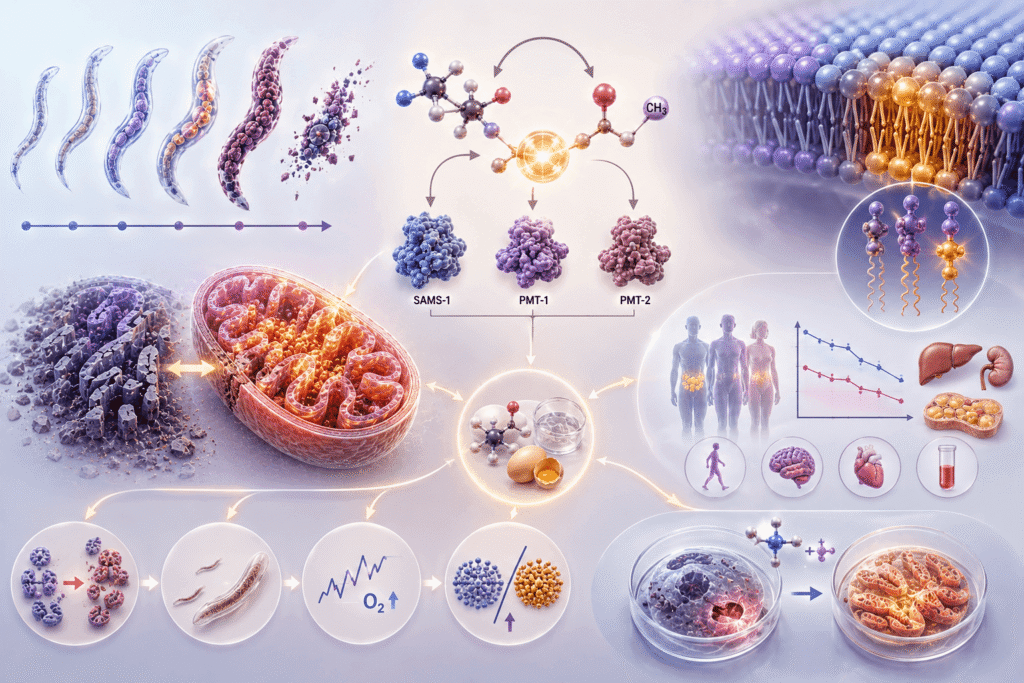
The study combines several model systems. The investigators began with longitudinal proteomics in wild-type C. elegans and two long-lived mitochondrial mutants, clk-1(qm30) and isp-1(qm150), sampled at young, post-reproductive, and old age. The mutants carry lifelong mitochondrial impairment yet live long, so they serve as a window into the adaptations that let an organism tolerate a struggling mitochondrion. A consistent finding emerged: metabolic and lipid-related changes were a late event in normal aging, whereas proteostasis and stress-response shifts came early.
Mining the proteome for the strongest, most progressive age-related losses returned S-adenosylmethionine synthetase (SAMS-1) as the top downregulated protein, and—critically—the phosphoethanolamine N-methyltransferases PMT-1 and PMT-2 as the next two. These three enzymes sit on the methylation arm of PC biosynthesis. Knocking down sams-1, pmt-1, or pmt-2 in young worms reproduced the mitochondrial picture of old age: a shift from tubular to fragmented and "very fragmented" mitochondria, reduced oxygen consumption rate on Seahorse respirometry, and reduced body size. Lipidomics confirmed the mechanism, showing reduced PC/PE and LPC/LPE ratios in the knockdowns.
The reversibility data are the heart of the paper. Supplementing PC directly, or choline (which feeds PC synthesis through the Kennedy pathway), rescued mitochondrial morphology, body size, respiration, and the lipid ratios in the knockdown animals—and, importantly, also alleviated the mitochondrial fragmentation and respiratory decline of normally aging wild-type worms. What’s crucial here is the speed and the timing: the correction was demonstrable in old animals, not only young ones, which is precisely what gives the finding clinical reach.
The authors then asked whether any of this translates. In the GTEx dataset, expression of PEMT—the human functional analog of PMT-1/2—declined with age across multiple tissues, most notably in lipid-rich organs such as subcutaneous and visceral adipose tissue and the ovary. In the UK Biobank NMR metabolomics data, total PC fell with age in men, while relative PC (PC normalized to total fatty acids) declined sharply in women after the approximate age of menopause, against a backdrop of rising total fatty acids. Higher PC and polyunsaturated fatty acid (PUFA) levels tracked with lower lactate, lower basal metabolic rate, lower comorbidity index, faster walking pace, and better memory—markers consistent with better mitochondrial function and healthier aging. Finally, in a human cell model, choline (best in combination with the Complex II substrate succinate) protected fibroblasts against metformin-induced loss of mitochondrial membrane potential, demonstrating conserved restoration of metabolic resilience.
The Mechanistic Chain
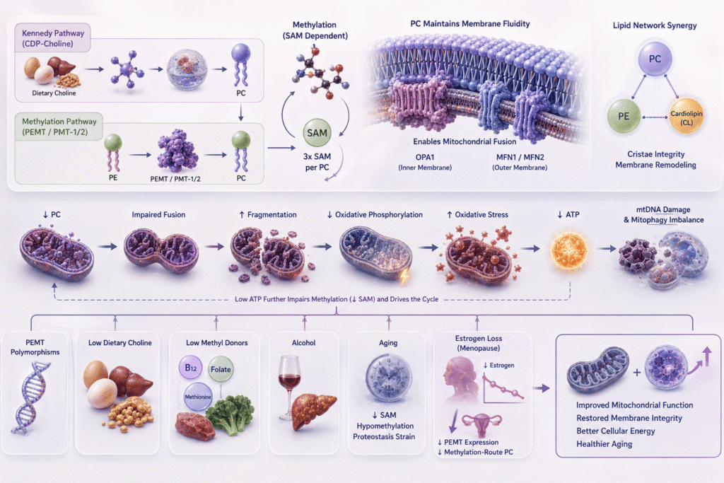
Based on my own synthesis (not a direct measurement made in the paper), let’s assemble the mechanism into a single conceptual chain. First, let’s look at the two routes to PC. The Kennedy (CDP-choline) pathway converts dietary choline to PC and runs in most tissues. The alternative route, the one the paper foregrounds, converts phosphatidylethanolamine (PE) to PC via methyltransferase activity—PEMT in humans, PMT-1/2 in worms—and this conversion requires methylation, with S-adenosylmethionine (SAM) as the methyl donor. What’s of real consequence here is that making a single PC molecule by this route consumes SAM three times, across three sequential methylation steps. That makes methylation-dependent PC synthesis an unusually heavy draw on the cell's methylation reserves, which is part of why it’s so vulnerable when SAM is limiting.
Now let’s review fusion. Mitochondrial fusion depends on a fluid, appropriately curved membrane, and PC—the most abundant phospholipid in both mitochondrial membranes—supplies that fluidity. Fusion involves OPA1 governing inner-membrane fusion, and Mitofusin-1 and Mitofusin-2 on the outer membrane. Looking beyond PC alone, it’s important to note that cardiolipin and PE participate in remodeling and crosstalk with PC to maintain cristae integrity—a point we’ve touched on for cardiolipin but not framed as a coordinated lipid network.
When PC declines, fusion fails, fragmentation rises, and the downstream consequences cascade: impaired oxidative phosphorylation, oxidative stress, reduced ATP, and—I argue—a self-reinforcing loop, because ATP is itself required for methylation. mtDNA failure and a mitophagy imbalance are further downstream features. The takeaway here is that this is not a single lesion but a connected collapse, seeded upstream by a lipid biosynthetic deficit.
Walking further upstream, let’s consider what reduces PC synthesis in the first place. The candidates include PEMT polymorphisms that blunt endogenous methylation-route synthesis; dietary insufficiency of choline (eggs, liver, soy) and of methylation cofactors such as B12, folate, and methionine; alcohol, which impairs hepatic PEMT; aging-related decline in SAM with consequent hypomethylation and proteostasis strain; and the loss of estrogen at menopause. It’s important to keep in mind that estrogen helps induce the PEMT enzyme, so its withdrawal at menopause removes a key driver of the methylation route, which I think of as the biological basis for the steep PC decline the Biobank data showed in women around that age.
The Clinical Translation
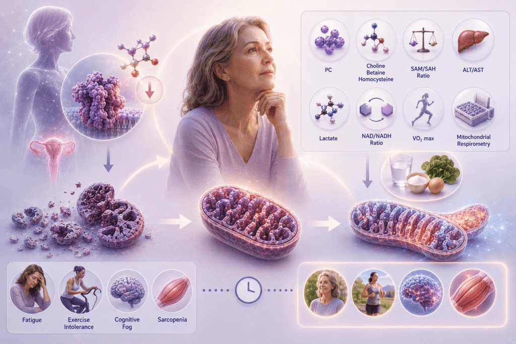
With all of this in mind, I propose reframing menopausal energy decline—the familiar cluster of fatigue, exercise intolerance, cognitive fog, and sarcopenia—not merely as "estrogen depletion and its effects on mitochondria," a framing that has long been used, but as a lipid-mitochondrial deficit. Estrogen still matters, but I believe it matters substantially because it gates a lipid pathway. I see this as a proposed, correlative model that makes transitional sense, not an established causal claim; indeed, the paper's own human data are explicitly correlative and descriptive.
This reframing can be paired with a practical biomarker map for clinicians wanting to watch this process: plasma phosphatidylcholine; choline, betaine, and homocysteine; the SAM-to-SAH ratio as a readout of methylation capacity; ALT and AST, since fatty liver disease degrades hepatic PEMT; lactate and the NAD/NADH ratio as indicators of the glycolytic shift that accompanies mitochondrial inefficiency; and functional measures such as VO2 max and direct mitochondrial respirometry (re mito-swab and Seahorse-type testing).
The most significant implication is the therapeutic window. Because the worm data show reversal in old animals, the intervention is not confined to the early-life paradigm that dominates much of aging research. The paper opens a distinct, later-life window for healthspan extension: nutritional intervention targeting PC can restore mitochondrial integrity "no matter where we are in this aging journey." For a clinician treating post-middle-age patients, that is the difference between prevention only and meaningful late-stage correction.
The Multi-Arm Strategy
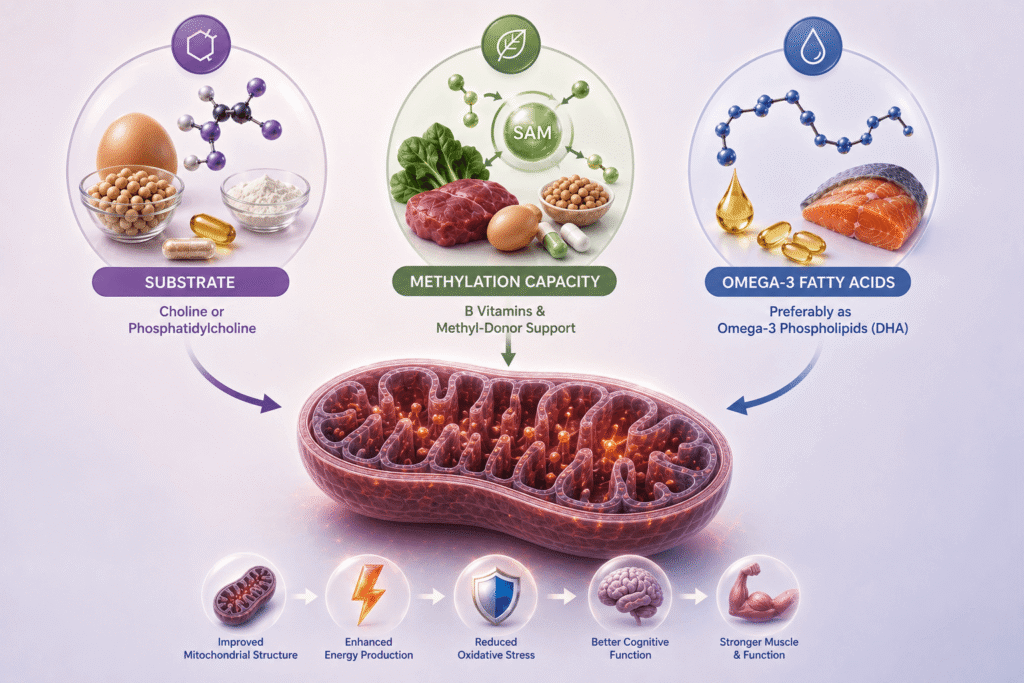
What I would like you all to carry forward is the idea that a single-substrate approach will not fully reverse a deficit of this kind. Repleting choline or PC alone, optimizing methylation alone, or adding omega-3s alone each addresses only one node of a connected system. I therefore advocate a three-arm clinical strategy:
- Substrate: choline or phosphatidylcholine directly, to feed PC production.
- Methylation capacity: the relevant B vitamins and methyl-donor support, recognizing how heavily the methylation route draws on SAM.
- Omega-3 fatty acids, preferably as omega-3 phospholipids: DHA is the fatty acid ideally incorporated into PC (a suggestion that the paper touches on), pairing with a precursor such as choline.
Why this pairing? The membrane being rebuilt is not generic PC but PC of the right composition for fluidity and curvature, and that the enzymatic machinery rebuilding it needs methyl donors to run. Address only one arm and the others remain rate-limiting.
Conclusion
I believe that the lasting contribution made in this paper is a reframing that mitochondrial aging is driven, in substantial part, by membrane lipids and an upstream biosynthetic deficit—not only by genetic decay or the other mechanisms the field usually invokes. PC synthesis declines progressively with age, most steeply in post-menopausal women, and that decline mechanistically links a phospholipid to whole-body fatigue and frailty in a way no prior study has validated and correlated so directly. That the deficit appears reversible, late in life, with accessible dietary tools is what makes it, in my view, a model of aging that is genuinely modifiable at the bedside—an upstream lever on the lipidome with real consequences for healthspan.
Reference:
Poliezhaieva, T., Li, Y., Chaudhari, P.S. et al. Aging-associated decline of phosphatidylcholine synthesis is a malleable trigger of natural mitochondrial aging. Nat Commun 17, 3589 (2026).
https://doi.org/10.1038/s41467-026-71508-7